Key Takeaway:
- Gram-positive bacteria are bacteria that appear purple after the Gram stain process, which is used in microbiology to distinguish them from Gram-negative bacteria.
- Gram-positive bacteria have a thick peptidoglycan layer in their cell walls, which is responsible for their purple color. They also have teichoic acid and lipoteichoic acid in their cell walls.
- Examples of Gram-positive bacteria include Bacillus, Staphylococcus, Streptococcus, Enterococcus, Listeria, Clostridium, Mycobacterium, Actinobacteria, and Firmicutes. Understanding the characteristics of Gram-positive bacteria is important in medical diagnosis and antibiotic treatment.
Understanding the Gram-staining process

Photo Credits: colorscombo.com by Eugene Hall
Gram-staining is a fundamental process in microbiology used to identify bacterial species. The unique characteristics of bacterial cells are highlighted by differential staining using the Gram’s method. By classifying bacterial cells into Gram-positive and Gram-negative groups, it is possible to distinguish their cell wall structural differences. The Gram-positive group appears purple under a microscope due to their thicker peptidoglycan layer, while the Gram-negative group appears red or pink due to their thin peptidoglycan layer. This method is essential in determining appropriate antibiotics to treat bacterial infections. To ensure accurate outcomes, it is crucial to follow correct procedures in Gram staining.
It is critical to note that the Gram-staining process includes four sequential steps: Fixation, staining with crystal violet, washing with an iodine solution, and finally decolorizing followed by counterstaining with safranin. The process should be handled with care, as any deviation in technique can affect the outcome. In diagnostics, the Gram-staining process allows for visual confirmation of bacterial cell morphology and their structural properties.
It is important to note that atypical bacteria may not adhere to the standard classification when using the Gram-stain method. Therefore, additional staining procedures should be employed to differentiate such bacteria.
Overall, understanding the Gram-staining process is vital in microbiology and early diagnosis of bacterial infections. Ensure that the procedure is correctly performed to obtain accurate outcomes.
What is Gram-positive bacteria?
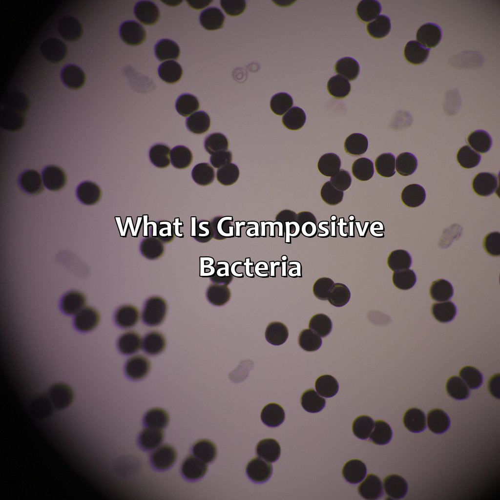
Photo Credits: colorscombo.com by Bradley Carter
Gram-positive bacteria are a type of bacteria that stains purple when exposed to the Gram stain test. This is because these bacteria have a thick peptidoglycan layer in their cell wall, which retains the crystal violet dye used in the test. They also contain teichoic acid and lipoteichoic acid which contribute to their bacterial morphology.
Gram-positive bacteria are responsible for causing various infections such as strep throat, pneumonia, and skin infections. A true fact is that the discovery of Gram-positive and Gram-negative types of bacteria is credited to Danish bacteriologist Hans Christian Gram in 1884.
Characteristics of Gram-positive bacteria
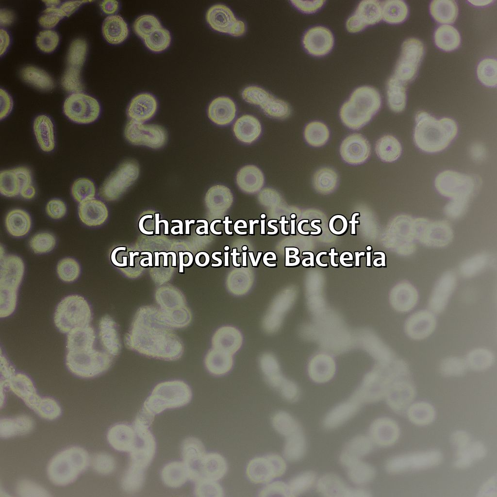
Photo Credits: colorscombo.com by Gabriel Clark
Unlocking the secrets of gram-positive bacteria? It’s all about their distinct cell membrane and wall structures. We’ll look at the “Characteristics of Gram-positive bacteria”. This includes how they react to Gram’s iodine and crystal violet staining. We’ll break it down into sections, including:
- Cell wall structure
- Membrane properties
- Shape/arrangement of Bacillus, Staphylococcus, Streptococcus, Enterococcus, Listeria, Clostridium, Mycobacterium, Actinobacteria, and Firmicutes
Cell wall structure
The cell wall structure of Gram-positive bacteria is unique and significant. The peptidoglycan layer forms the primary component of the cell wall, which provides rigidity and structural integrity. In addition, teichoic acid and lipoteichoic acid are essential components that give a negative charge to the cell surface while also function as receptors for bacteriophages and antibiotics. Further, teichoic acid plays a role in the secretion of virulence factors and serves as an anchor for various proteins on the bacterial surface.
A key feature of the cell wall in Gram-positive bacteria is its ability to retain crystal violet during staining due to its thick layer of peptidoglycan. The retention of dye results in these organisms appearing purple under a microscope after the staining process.
A pro tip for identifying Gram-positive bacteria based on their cell wall structure is to examine their sensitivity to antibiotics such as penicillin. Gram-positive bacteria with thick peptidoglycan layers are generally more susceptible to this class of antibiotics, making them easy to treat compared to Gram-negative bacteria with thinner peptidoglycan layers.
Membrane properties may not sound sexy, but for Gram-positive bacteria they’re essential for survival.
Membrane properties
The cell membrane is an important aspect of the Gram-positive bacteria. This thin, delicate layer is responsible for separating and protecting the bacterial cell from its surroundings while allowing nutrients in and waste out.
| Cell Membrane Structure | Composed of a phospholipid bilayer with embedded proteins and teichoic acids |
| Periplasmic Space | Small periplasmic space between the cytoplasmic membrane and peptidoglycan layer |
| Variability | The thickness and lipid composition of Gram-positive cell membranes can vary among different species. |
Interestingly, some antibiotics, such as polymyxin, take advantage of the cell membrane’s vulnerability to disrupt or lyse (break apart) Gram-positive bacteria.
Pro Tip: Understanding the structure and function of the cell membrane in bacteria can help identify suitable antibiotic treatments.
A bacterial world of rods and spheres, where morphology is king and Firmicutes reign supreme.
Shape and arrangement
Gram-positive bacteria exhibit diverse bacterial morphology, ranging from cocci (spherical) to bacillus (rod-shaped), and can grow in different arrangements. Some common shapes include single cells, clusters, chains, and pairs.
| Shape | Example |
|---|---|
| Cocci | Staphylococcus aureus, Streptococcus pneumoniae, Enterococcus faecalis |
| Bacillus | Listeria monocytogenes, Clostridium difficile |
| Mycobacterium | Mycobacterium tuberculosis |
| Actinobacteria | Corynebacterium diphtheriae |
| Firmicutes | Staphylococcus epidermidis, Streptococcus pyogenes |
Interestingly, Bacillus can also produce spores that enable them to survive extreme environments such as high temperatures or low pH levels.
The different shapes and arrangements of Gram-positive bacteria are a result of how they divide during cell growth and the structure of their cell walls. These unique features allow for effective diagnosis and treatment of bacterial infections. However, some bacteria such as Clostridium difficile can cause severe illnesses like colitis in humans.
Staining Gram-positive bacteria is like a purple party – where crystal violet dances with safranin and everyone is left feeling positively purple.
Gram-positive staining process
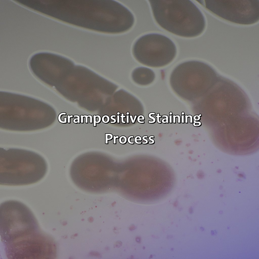
Photo Credits: colorscombo.com by Brandon Brown
Gram stain is a method devised by Hans Christian Gram to understand the Gram-positive staining process in microbiology. It involves four steps: crystal violet, safranin, decolorization, and counterstain. This section will discuss the mechanism of Gram-positive staining. Plus, we’ll explore the steps included in Gram-positive staining using the differential staining technique of Gram’s method.
Steps involved in Gram-positive staining
The Gram staining method is a crucial process in microbiology for identifying the characteristics of bacteria. To perform the Gram-positive staining, first, clean and make a bacterial smear on a glass slide. Second, allow it to air dry to prevent excess moisture. Third, heat fixation is performed to allow attachment of bacteria to glass slides. Fourth, the Gram’s method is applied and rinsed using decolorizer.
- Clean and make a smear of bacteria on a slide
- Air-dry the slide
- Heat fixation of smear
- Apply Gram’s method and rinse using decolorizer
Applying the dye crystal violet to bind with peptidoglycan layers creates a purple color on cells’ surface; lastly, they are counterstained with safranin to give them their distinct red coloration under light microscopy.
The uniqueness of each stained bacterium helps in identification as different bacteria display different cell composition and physiology. In general, this process proves useful in differentiating two types of seminal bacterial groups, Gram-Positive (violet) and Gram-Negative (pink). It is worth noting that cell wall differentiation takes place due to differences in thickness as well as chemical composition.
According to the Royal Microscopical Society’s research “Biology: A Global Approach,” “Following microscopic observations which revealed that infectious microbes typically grouped into two main types depending on reaction outcome after treatment with certain dyes – one group dyeing blue [Gram positive] whereas other rod-shaped organisms did not take up the stain,” which implies significance in understanding gram stains’ importance.
Making bacteria look pretty under the microscope with the Gram stain method is like giving them a full makeover.
Mechanism of Gram-positive staining
Gram-positive staining is a critical process in the field of microbiology. It involves differential staining of bacterial cells to identify their type based on their cell wall properties. As Gram-negative bacteria have an outer membrane, whereas Gram-positive bacteria do not, they react differently during this process. The mechanism of Gram-positive staining involves applying crystal violet dye followed by iodine and alcohol washes. Due to the thick peptidoglycan layer in their cell wall, the dye gets trapped within Gram-positive bacteria, resulting in a purple coloration upon microscopic examination. This unique property allows for easy differentiation and identification of bacterial cells.
In addition to the dyes used in the process, several factors affect the mechanism of Gram-positive staining, including the duration and intensity of exposure to various chemicals used in the method. For instance, exposing a Gram-stained slide to too much alcohol may cause loss of dye from both types of cells leading to an incorrect diagnosis. Therefore using controlled conditions for appropriate soaking times are crucial as well as preparation of bacterial cells through drying and heat fixing process before gram’s method application ensures better sensitivity.
The significance of understanding the mechanism behind Gram-positive staining lies primarily in its widespread usage across microbiology labs worldwide for patient sample analysis, antibiotic susceptibility testing; it aids clinicians with better treatment approach accordingly.
With this knowledge in mind, microbiologists can accurately differentiate between various bacterial species based on their color and structure under illumination. Furthermore, recognizing different bacterial species allow for better understanding their pathogenicity levels- one capability that has significant consequences for public health welfare against zoonotic infections such as deadly pandemic diseases like COVID-19 caused by SARS-CoV-2 which still need robust research on testing rapidly and accurately where gram stain technique adopted right off-the-bat could save headlines!
Gram-positive bacteria may be purple under the microscope, but they’re not as regal as they seem – just ask the microbiologist who stains them.
Color and appearance of Gram-positive bacteria

Photo Credits: colorscombo.com by Larry Mitchell
Gram-positive bacteria exhibit a purple color when stained with crystal violet. Safranin is used as a counterstain to color gram-negative bacteria pink. Examining staining properties closely is the solution to understand the color and appearance of these bacteria.
Microbiology studies the primary and counter staining techniques, and the appearance of these bacteria under a microscope.
Primary stain
The primary stain is the first step in Gram-positive staining, which uses a purple dye called crystal violet to colorize bacteria. It is an essential step in differentiating between Gram-positive and Gram-negative bacteria.
| Primary Stain | Description | Example |
|---|---|---|
| Purple Dye (Crystal Violet) | A basic stain that binds to the peptidoglycan layer of the cell wall. | Bacillus anthracis |
Gram-positive bacteria have a thicker cell wall that retains crystal violet during the staining process, resulting in a purple coloration. This initial step highlights the bacteria’s cell morphology, and it helps differentiate it from other types of bacteria.
It is crucial to ensure that proper staining techniques are followed when working with Gram-positive bacteria as misidentification of bacteria could lead to inaccurate diagnosis, treatment, or even death.
Knowing how to distinguish between different types of bacterial cells can help prevent the spread of infectious diseases. Ensuring accurate identification of pathogenic organisms could also play an important role in preventing outbreaks and pandemics. Stay informed!
Even after staining purple with crystal violet, Gram-positive bacteria are still thirsty for attention and require a splash of safranin to really stand out under the microscope.
Counter stain
A counterstain is an essential component of the Gram-staining process used to differentiate between Gram-positive and Gram-negative bacteria. After applying the primary stain, crystal violet, which highlights all bacteria in a purple shade, a decolorizer solution removes the stain from the outer membrane layer of Gram-negative bacteria while leaving behind stains on the thicker peptidoglycan cell wall of Gram-positive bacteria. At this point, the counterstain, safranin, is applied to all cells that have lost their primary stain to highlight their presence under a microscope. Counterstaining allows us to differentiate between purple-stained Gram-positive bacteria and red-pink stained Gram-negative bacteria.
Unique details can be mentioned about how counter staining works and why safranin is used for this process. Safranin-which stains cell material such as nucleic acid and protein in shades of red – has a low affinity for crystal violet that has already stained the thick peptidoglycan cell walls of Gram-positive cells. The counterstain instead enters through the thinner outer membrane of gram-negative bacteria which have lost their primary stain due to decolonization but have yet retained some residue of it at certain areas like Endospore.
Some suggestions include:
- Using a mordant (iodine) before decolorizing to prevent loss of primary stain due to its removal by alcohol or acetone.
- Always using fresh bacterial cultures with distinct color differences even though over time some strains can lose their affinity for certain stains.
- Performing quality control checks for accuracy purposes like comparing unstained and stained preparations on different microscopes and slide batches.
Counter staining remains one of the most crucial aspects of gram-positive vs. gram-negative identification under a microscope with better stability against erroneous identification labels between these two categories.
Get ready to zoom in and witness the colorful world of Gram-positive bacteria under the microscope.
Appearance under the microscope
The appearance of Gram-positive bacteria under the microscope reveals distinct characteristics that assist in their identification and diagnosis. The staining process reveals vivid colors, which allow microbiologists to differentiate between different bacterial types.
Upon observing Gram-positive bacteria under the microscope, their cell walls appear dark blue or purple due to the absorption of crystal violet dye during the staining process. The coloration is a result of the thick peptidoglycan layer in their cell wall structure, which provides rigidity and protection for the cell.
The shape and arrangement of Gram-positive bacteria can also be observed under the microscope. Some may appear as spherical clusters (Staphylococcus), while others may form chains (Streptococcus) or rods (Clostridium).
Additional details can also be observed through acid-fast staining techniques and decolorization processes. These techniques help microbiologists distinguish between different types of bacteria based on their unique properties.
To ensure accurate observations and analysis, it is important to use a properly calibrated microscope, maintain sterile conditions throughout staining procedures, and adhere to established protocols for handling and analyzing bacterial samples.
Gram-positive bacteria: the good, the bad, and the ugly of medical microbiology.
Importance of Gram-positive bacteria

Photo Credits: colorscombo.com by Stephen Torres
Gram-positive bacteria can have both positive and negative influences on the human body. Probiotics and prebiotics can be beneficial. However, bacterial resistance, sepsis, and various infections caused by community and healthcare-associated pathogens, plasmid transfer, and gene mutation can be dangerous. To understand the importance of gram-positive bacteria in medical diagnosis and bacterial infections, it’s important to know both effects.
Positive effects
Positive Effects:
Gram-positive bacteria have been found to have several beneficial effects on the human gut and the environment. These effects include:
- A stabilizing effect on the microbial population
- Improved nutrient absorption and digestion
- Prevention of harmful bacterial colonization
Gram-positive bacteria are also used in the production of probiotics and prebiotics, which help support digestive health and boost immunity. Additionally, some gram-positive bacteria are used in cheese production and other food processing applications.
It is interesting to note that certain species of gram-positive bacteria have also been found to produce antimicrobial peptides that can inhibit the growth of harmful bacteria. This research was conducted by scientists at Boston Children’s Hospital.
Gram-positive bacteria may have a sunny disposition, but their negative effects can lead to medical mayhem and antibiotic nightmares.
Negative effects
The Harmful Effects of Gram-positive Bacteria
Gram-positive bacteria can have negative effects on medical diagnosis and treatment by causing bacterial infections that can lead to severe conditions such as sepsis. The use of antibiotics such as penicillin, vancomycin, rifampin, erythromycin, tetracycline, and ciprofloxacin has been widely used to treat these infections. However, bacterial resistance to these antibiotics has also become a growing problem in recent years.
Bacterial infections caused by Gram-positive bacteria are commonly observed in community-acquired infections and healthcare-associated infections due to their ability to cause severe diseases like bacterial pneumonia, skin infections, bloodstream infections, endocarditis, urinary tract infections, foodborne illness, diarrhea and toxic shock syndrome. These nosocomial infections in particular can hinder patient recovery as they tend to occur in individuals already undergoing medical treatment.
The transfer of plasmids during horizontal gene transfer has led to increased genetic diversity within the species of Gram-positive bacteria leading to antibiotic resistance development. Gene mutations provide further ways for these bacteria to evolve and become resistant.
When it comes to dealing with Gram-positive bacteria, knowing your antibiotics is the difference between a medical diagnosis and a bacterial infection.
Examples of Gram-positive bacteria
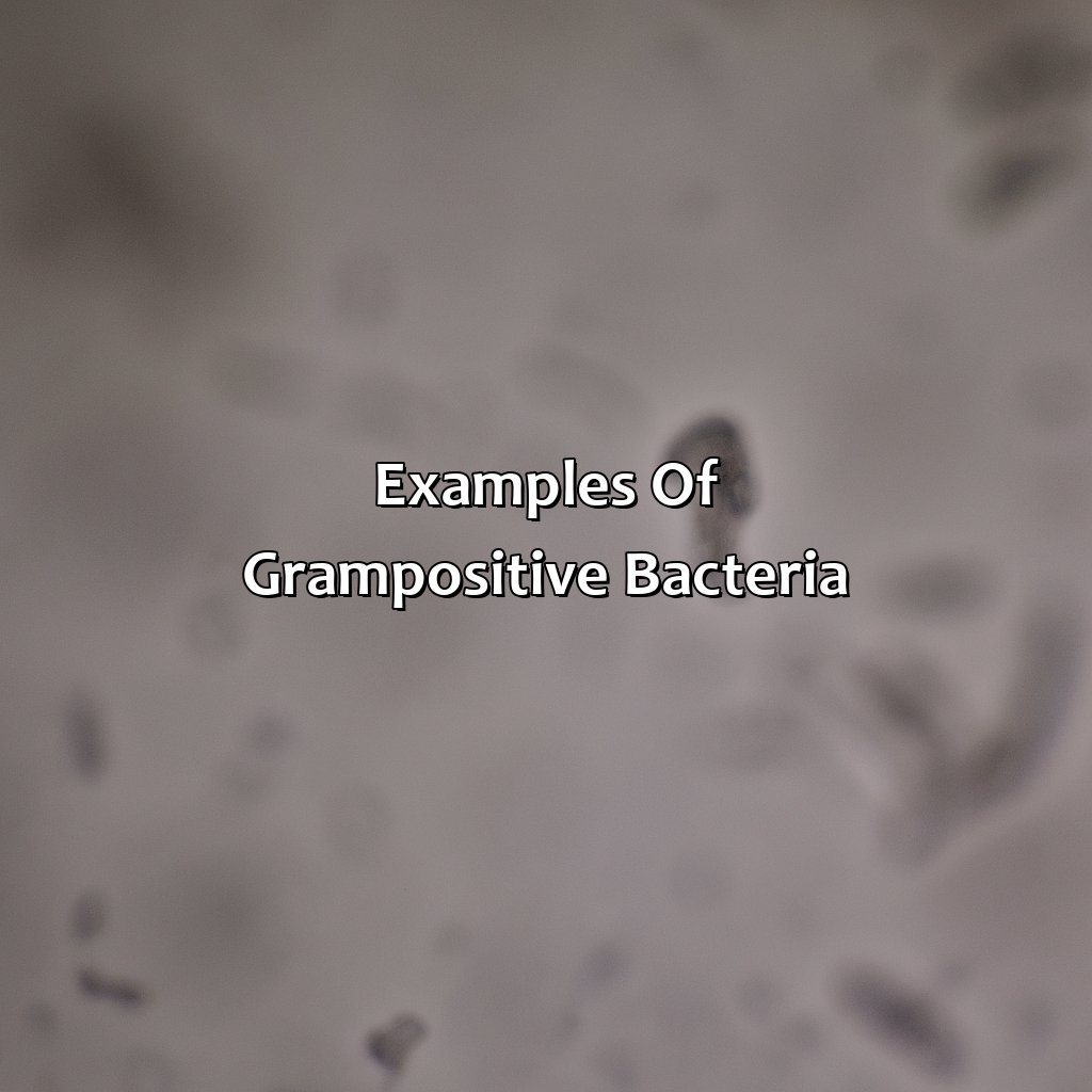
Photo Credits: colorscombo.com by Roger Gonzalez
Want to know more about gram-positive bacteria and medical diagnosis? Check out our examples section! We’ll explore Staphylococcus aureus, which can cause community-acquired and healthcare-associated infections. These include bacterial pneumonia, skin and bloodstream infections. Then there’s Streptococcus pneumoniae, which can lead to sepsis and requires penicillin treatment. Finally, Clostridium difficile is linked to bacterial infections, sepsis, diarrhea and nosocomial infections. It is treated with vancomycin.
Staphylococcus aureus
This Gram-positive bacterium, often abbreviated as S. aureus, is a common cause of both community-acquired and healthcare-associated infections. It has the ability to cause a range of diseases including bacterial pneumonia, skin infections, bloodstream infections, and endocarditis. MRSA is a type of S. aureus that is resistant to many antibiotics and can be particularly difficult to treat.
S. aureus is known for its ability to produce toxins that can contribute to the development of disease. It also has unique cell surface structures that allow it to evade the immune system and thrive in different environments.
Interestingly, S. aureus was once associated mainly with hospital settings but has now become increasingly common in the community at large. This shift has led to increased efforts to identify and control the spread of MRSA, particularly in high-risk populations such as those in healthcare facilities or with compromised immune systems.
Despite its negative impact on human health, S. aureus also plays an important role in our natural microbiome and has been found on healthy human skin and mucous membranes. Understanding the complex interactions between humans and bacteria such as S. aureus remains an area of active research with implications for both prevention and treatment of bacterial infections.
Streptococcus pneumoniae may sound like a fancy Italian dish, but it’s actually a gram-positive bacteria responsible for nasty infections like pneumonia and sepsis.
Streptococcus pneumoniae
Streptococcus pneumoniae, commonly referred to as pneumococcus, is a bacteria that belongs to the Gram-positive group. It has a distinctive morphology of small, spherical cells that usually form chains. Pneumococcus is responsible for causing a range of infections such as:
- bacterial pneumonia
- sinusitis
- otitis media and meningitis
In medical diagnosis, identifying whether an infection is caused by pneumococcus or other bacteria is essential for effective antibiotic treatment. Due to its high incidence in community-acquired infections and healthcare-associated infections, pneumococcus poses substantial health risks and can result in sepsis if left untreated. Interestingly enough, penicillin has been utilized since its discovery in 1928 to treat pneumococcal infections. Despite resistance issues arising against penicillin and other antibiotics over the years, pharmaceuticals continue efforts to develop new treatments against Streptococcus pneumoniae.
Why worry about a pandemic when you can have a nosocomial infection of Clostridium difficile?
Clostridium difficile
Clostridium difficile is a gram-positive, anaerobic, spore-forming bacterium associated with antibiotic-associated diarrhea and other infections. Its diagnosis relies on the detection of toxins A and B in stool specimens using specialized medical tests. C. difficile infections are commonly found in hospitals and healthcare facilities as its spores can live on surfaces for long periods, making it a primary cause of nosocomial infections.
Antibiotic treatment is usually recommended for severe cases, such as metronidazole or vancomycin. However, some strains have become resistant to these treatments, resulting in much more severe outcomes such as sepsis or even death.
Five Facts About Gram Positive Bacteria:
- ✅ Gram positive bacteria have a thick cell wall composed of peptidoglycan. (Source: Microbiology Society)
- ✅ Gram positive bacteria stain purple or blue when subjected to the Gram staining method. (Source: TeachMe)
- ✅ Some common examples of Gram positive bacteria include Staphylococcus and Streptococcus species. (Source: Healthline)
- ✅ Gram positive bacteria can cause illnesses such as meningitis, pneumonia, and sepsis. (Source: Centers for Disease Control and Prevention)
- ✅ Many antibiotics, such as penicillin, target Gram positive bacteria by interfering with their cell wall synthesis. (Source: Microbiology and Molecular Biology Reviews)
FAQs about Gram Positive Is What Color
What color is gram positive bacteria?
Gram positive bacteria appear purple or blue when stained with crystal violet and iodine.
How can gram positive bacteria be identified?
Gram positive bacteria can be identified through a staining process known as the Gram stain, which involves using crystal violet, iodine, alcohol, and safranin.
What is the cell wall of gram positive bacteria made of?
The cell wall of gram positive bacteria is composed of a thick layer of peptidoglycan, teichoic acids, and lipoteichoic acids.
What types of infections are caused by gram positive bacteria?
Gram positive bacteria can cause a range of infections, including skin infections, respiratory tract infections, and bloodstream infections.
Are all bacteria that are purple gram positive?
No, not all purple bacteria are gram-positive. Some bacteria may appear purple due to other staining methods or pigments.
What is the difference between gram positive and gram negative bacteria?
The main difference between gram positive and gram negative bacteria is in the composition of their cell walls. Gram positive bacteria have a thick layer of peptidoglycan in their cell walls, while gram negative bacteria have a thin layer of peptidoglycan surrounded by an outer membrane containing lipopolysaccharides.