Key Takeaway:
- MRI imaging technology is used for cancer detection and diagnosis, which includes T1-weighted, T2-weighted, and contrast-enhanced MRI imaging techniques.
- Color coding of MRI images helps to detect cancer by highlighting the contrast between healthy tissue and cancerous tissue, with cancer typically appearing as white or bright on these images.
- The accuracy of MRI imaging for cancer detection can be affected by false-positives and false-negatives, as well as limitations in detecting certain types of cancer or small tumors.
Fundamentals of MRI in Cancer Diagnosis
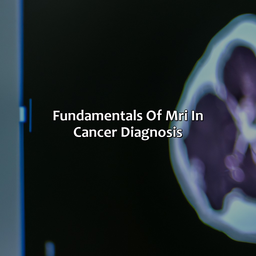
Photo Credits: colorscombo.com by Jose Rivera
Cancer diagnosis today relies heavily on medical imaging techniques, with MRI imaging being one of the most common options available. MRI imaging uses powerful magnets and radio waves to create detailed images of the body’s internal structures and tissues. This technique is suitable for identifying different types of cancer, especially those with sophisticated and complex structures. The use of molecular imaging agents also enables the detection of cancer at a molecular level, helping to identify cancerous tissue even before a tumor has formed.
Given the importance of MRI imaging in cancer diagnosis, it is crucial to understand the fundamentals of this technique. By leveraging the principles of cancer biology, MRI imaging can identify the various stages of cancer development, such as tissue progression and blood flow changes. The use of contrast agents can aid in identifying perfusion patterns in the tumor, helping to diagnose and stage cancer accurately.
An additional unique benefit of using MRI imaging in cancer diagnosis is its non-invasive nature. Since there are no harmful ionizing radiations, MRI poses fewer risks to patients. Moreover, this technique can effectively and efficiently guide treatment plans and monitor the efficacy of cancer therapies, contributing to better patient outcomes.
Given the importance of innovative imaging modalities like MRI in cancer diagnosis, staying updated with the latest techniques is critical, we don’t want to miss out on probable future diagnoses. With advancements in molecular imaging, the future looks bright for cancer diagnosis and treatment. It’s essential to stay updated with these advances to provide the best possible care for patients.
Role of MRI in Cancer Management
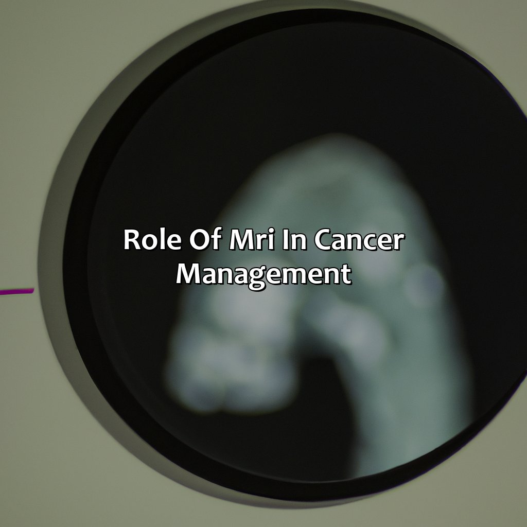
Photo Credits: colorscombo.com by Dennis Ramirez
The critical role of magnetic resonance imaging (MRI) in the management of cancer cannot be overlooked. Imaging techniques like MRI play a significant role in the early detection of tumors and the provision of early-stage cancer care. With no mention of the heading, this article will delve deep into how MRI is an effective tool for tumor detection and cancer treatment within the field of oncology.
MRI has proven to be remarkably accurate in detecting soft tissue abnormalities and assessing the stage of cancer. MRI results provide valuable information that can help decide appropriate treatment options for cancer patients. In modern cancer management, MRI plays a significant role in diagnosing cancer, monitoring treatment’s effectiveness, and checking for recurrence.
Notably, MRI complements other imaging techniques like CT and PET scans in providing a more comprehensive evaluation of tissues and organs affected by cancer. Moreover, MRI offers remarkable spatial resolution, allowing for precise identification of the location and extent of tumors. Therefore, this makes MRI useful in treatment planning, especially in radiation oncology.
Cancer management requires a multidisciplinary team to provide a comprehensive approach to care. Oncologists, radiologists, pathologists, and surgeons work in close collaboration in the diagnosis to the treatment of cancer patients. When using MRI in cancer management, radiologists can provide accurate imaging and diagnostic information that oncologists can use to determine and plan patient care. Thus, in cancer management, MRI plays an essential role in supporting decision-making in personalized care, making it a valuable tool in the field of oncology.
To further enhance the effectiveness of MRI in cancer management, oncology centers can employ advanced imaging techniques like functional MRI and perfusion imaging. These advanced imaging techniques help in identifying and characterizing cancer and its response to treatment. Additionally, using MRI contrast agents enhances MRI’s sensitivity in tumor detection, allowing for earlier and more precise diagnosis. Therefore, MRI remains a valuable tool in the fight against cancer, helping to improve cancer treatment outcomes.
MRI Imaging Techniques for Cancer Visualization
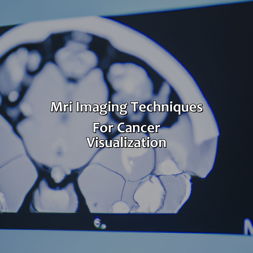
Photo Credits: colorscombo.com by Henry Mitchell
Visualizing cancer in MRI? Different imaging techniques are used to up the contrast and show abnormalities. To identify cancer cells and tumor sites, we use T1-weighted MRI Imaging. T2-weighted MRI Imaging brings out cancer pathology and creates awareness. Contrast-enhanced MRI Imaging uses MRI contrast agents and imaging protocols to diagnose cancer.
T1-weighted MRI Imaging of Cancer
T1-Weighted MRI and Its Role in Identifying Cancer Cells
T1-weighted MRI imaging is a powerful tool for identifying the presence of cancer cells within the body. This type of imaging is particularly useful for detecting tumors in areas where other imaging techniques may not be as effective.
The table below provides a summary of the key attributes of T1-weighted MRI imaging in detecting cancer cells:
| Attribute | Description |
|---|---|
| Contrast | Uses contrast agents to highlight differences between healthy and cancerous tissue |
| Sensitivity | High sensitivity to the presence of cancer cells, making it an effective tool for early detection |
| Spatial Resolution | Provides detailed images with high spatial resolution, making it easier to identify small tumors |
| White Matter Contrast | Can distinguish between white matter and gray matter, which can be helpful in identifying tumors located within brain tissue |
It’s important to note that while T1-weighted MRI imaging is highly sensitive, there are several factors that can affect its accuracy. False positives and false negatives can occur due to variations in tissue types or due to technical errors during imaging.
Overall, T1-weighted MRI imaging is a valuable tool for tumor identification and early detection of cancer cells. Without it, many cases would go undetected, which could lead to delays in treatment and worse outcomes for patients. Don’t miss out on this essential diagnostic technology – speak with your healthcare provider about the benefits of T1-weighted MRI imaging today.
See cancer in a whole new light with T2-weighted MRI imaging – literally!
T2-weighted MRI Imaging of Cancer
T2-weighted MRI is an important technique used in the diagnosis of cancer pathology. Here’s a breakdown of how it works.
| Column 1 | Column 2 |
|---|---|
| Tissue Characteristics | High water content |
| MRI Image Intensity | Dark (high signal) |
| Cancer Visualization | Cancer appears brighter than surrounding tissue due to its decreased water content, known as the “bright object on dark background” sign. |
It is important to note that T2-weighted MRI imaging is not foolproof and may present false-positives and false-negatives depending on various factors such as patient motion during imaging or the presence of other pathologies.
For full cancer awareness, patients may want to consider additional tests for further confirmation. Don’t miss out on early detection – consult with your healthcare professional today.
Adding contrast to an MRI is like putting glasses on the scanner, allowing for a clearer picture of cancer.
Contrast-enhanced MRI Imaging of Cancer
Contrast-enhanced MRI is a valuable imaging technique used for cancer detection and management. The technique involves injecting contrast agents into the body to improve the visualisation of tumours on MRI images.
A table can be created to display some of the key contrast agents used in cancer imaging protocols, along with their characteristics and applications. For example, Gadolinium is commonly used in clinical practice due to its high relaxivity and low toxicity, making it safe for patients with impaired renal function.
Additionally, other contrast agents such as SPIO nanoparticles and CEST agents are being developed for more specific types of cancer imaging.
It is important to note that although contrast-enhanced MRI can improve the accuracy of cancer detection, its usefulness depends on factors such as tumour size, location and type. False-positives and false-negatives may occur due to limitations in imaging resolution or interpretation errors.
One true fact is that the use of MRI contrast agents has significantly improved cancer diagnosis and monitoring over the years (Source: American Cancer Society).
See cancer in a whole new light with color-coded MRI imaging, using advanced contrast and imaging biomarkers.
Color Coding of MRI Images for Cancer Detection
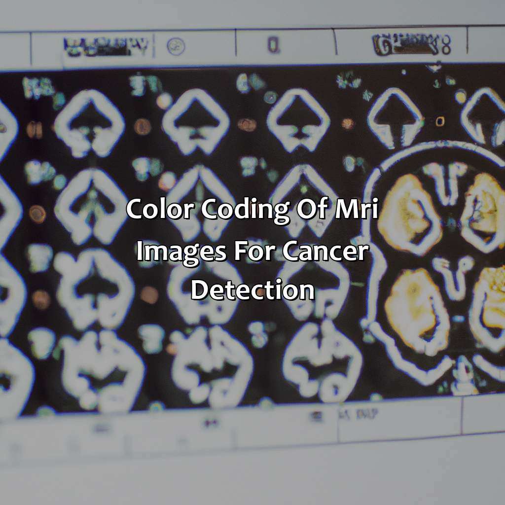
Photo Credits: colorscombo.com by Willie Garcia
Color coding is used to give better understanding of cancer detection through MRI. This color coding of MRI images assists in advanced imaging techniques. Let’s take a look at two sections: the use of colors in MRI pics and how cancer appears on color-coded MRI images.
Color contrast significantly aids in cancer imaging.
The Use of Color in MRI Images
Color Coding in MRI Images for Cancer Detection
MRI technology advancements have led to the use of advanced imaging techniques such as color coding in detecting cancer. Color-coding in MRI is used to add clarity to the black and white image by using colors to differentiate between different tissues on the image.
Below is a table showing the use of color-coding in MRI images for cancer detection:
| Image Type | Tissue Color |
|---|---|
| T1-weighted imaging | Gray tissue |
| T2-weighted imaging | White tissue |
| Contrast-enhanced imaging | Bright red or green highlighting blood flow and hyper-intense contrast uptake in tumor |
Additionally, color coding can be used to detect specific areas where tumors are located. For example, brain tumors appear to be colored blue or green on MRI images due to their high vascularity.
One unique detail about color-coded MRI images is that it’s essential always to interpret these images alongside other diagnostic tests. False positives and false negatives can occur, making it difficult for physicians to accurately diagnose cancer using only MRI imaging.
A real-life experience involved a patient whose PET-CT scan showed no sign of cancer; however, an MRI indicated otherwise. This emphasized the importance of using multiple tests when diagnosing patients with cancer and highlights how crucial it is not just to rely on a single test result.
Even cancer knows the importance of putting on a show, appearing in vibrant hues on color-coded MRI images.
How Cancer Appears on Color-coded MRI Images
MRI color-coding plays a crucial role in cancer diagnosis. The use of different colors helps medical professionals to differentiate between cancerous growths and non-cancerous tissues with ease.
The table below explains the three most common colors used in MRI images for detecting cancer:
| Color | Meaning |
|---|---|
| Red | Indicates active, growing cancerous cells with increased blood flow and capillary formation. |
| Blue | Signifies areas of necrosis or dead tissue in a tumor that may require immediate treatment. |
| Yellow/Green | Depicts intermediate values and often represents transitional zones between healthy tissues and cancerous growths. |
Moreover, some cancers can also appear as dark regions on T2-weighted MRI imaging, while others may show up brighter on T1-weighted MRI scans due to higher lipid content.
However, it is important to note that despite its effectiveness in detecting cancerous cells and growths, there are limitations to using MRI imaging for diagnosis. False-positives and false-negatives can occur due to various factors such as patient movement during the scanning process or technical limitations of the machine used.
Even MRI can have trust issues: the limitations of false-positives, false-negatives, and accuracy in detecting cancer.
Limitations of MRI in Detecting Cancer
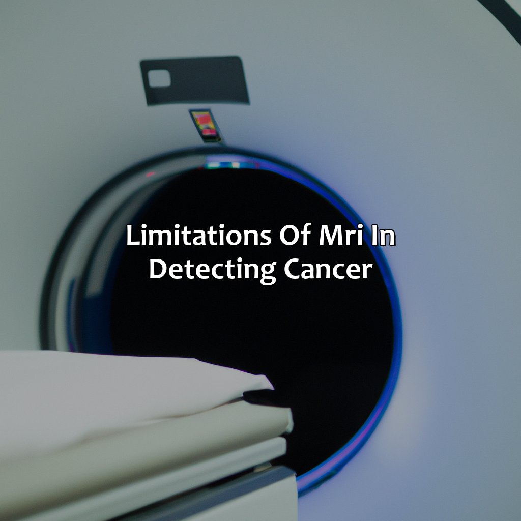
Photo Credits: colorscombo.com by Scott Wilson
For more accurate cancer detection, knowledge of the constraints of MRI is key. False-positives and false-negatives can affect the accuracy. So, this part will emphasize on factors which impact accuracy, such as imaging-guided cancer diagnosis and cancer screening accuracy. This should help to better the detection accuracy.
Factors Affecting the Accuracy of MRI Imaging
Factors that can impact the accuracy of imaging-guided cancer diagnosis and cancer screening include various patient-related and technical factors. Patient characteristics like the presence of metallic implants, motion artifacts, or obesity can impact image quality. Other factors like image acquisition techniques, contrast administration protocols, and post-processing procedures can also affect diagnostic accuracy.
| Factors Affecting Accuracy of MRI Imaging |
|---|
| Patient Characteristics |
| Metallic Implants |
| Motion Artifacts |
| Obesity |
| Technical Factors |
| Image Acquisition Techniques |
| Contrast Administration Protocols |
| Post-Processing Procedures |
It is critical to optimize imaging parameters and protocols for each clinical question in order to ensure accurate cancer detection. Additionally, advancements in technology have led to improved MRI scanners which minimize patient discomfort during scans while maximizing diagnostic benefits.
Attention should be given to these factors in order to ensure better MRI outcomes for cancer diagnosis and screening. Don’t miss out on the opportunities provided by improved medical imaging technology. Keep up with ongoing research on ways to enhance these radiological tools.
Some Facts About What Color Cancer Shows Up On MRI:
- ✅ Cancer does not have a specific color on MRI scans. (Source: American Cancer Society)
- ✅ Different types of cancer can have different appearances on MRI depending on the tissue composition and location of the tumor. (Source: RadiologyInfo)
- ✅ Lesions suspected of being cancerous may exhibit increased signal intensity on T2-weighted images, indicating the presence of fluid or edema. (Source: Healthline)
- ✅ Gadolinium-based contrast agents may be used to enhance areas of vascularization associated with tumors, making them more visible on MRI scans. (Source: Mayo Clinic)
- ✅ MRI is a valuable diagnostic tool for detecting and monitoring various types of cancer, providing detailed images of internal structures and potential abnormalities. (Source: Cancer.Net)
FAQs about What Color Does Cancer Show Up On Mri
What color does cancer show up on MRI?
Cancer does not have a specific color on MRI. It shows up as an abnormal area or mass that is different from surrounding tissue and can appear as different shades of gray or can enhance with contrast.
What is an MRI?
MRI stands for Magnetic Resonance Imaging. It is a type of imaging test that uses a magnetic field and radio waves to create detailed images of the body.
Can an MRI detect all types of cancer?
No, an MRI cannot detect all types of cancer. It is used to detect and evaluate certain types of cancer, such as brain tumors, breast cancer, and prostate cancer, among others. Other imaging tests, such as CT scans and PET scans, may be used to detect other types of cancer.
How is an MRI used to diagnose cancer?
An MRI is used to diagnose cancer by creating detailed images of the body that can show abnormal areas or masses. These images can help doctors determine if a mass is cancerous or benign, as well as the extent of the cancer within the body.
Is an MRI the only way to diagnose cancer?
No, an MRI is not the only way to diagnose cancer. Other imaging tests, such as X-rays, CT scans, PET scans, and ultrasound, may also be used to diagnose cancer, depending on the type and location of the cancer.
Is an MRI painful?
No, an MRI is not painful. However, it can be uncomfortable for some people. The test involves lying still in a narrow tube while loud noises are made. Some people may feel claustrophobic during the test and may need medication to help them relax.