
Key Takeaways:
- Pelvic ultrasound plays a major role in the early detection and diagnosis of cancer, particularly gynecological cancer. This minimally invasive imaging technique can help identify abnormal growths, masses, and tissues in the pelvic area.
- Understanding pelvic ultrasound requires knowledge of ultrasound technology, tumor imaging, pelvic anatomy, and color coding in ultrasound. Different colors on the ultrasound image represent different phenomena such as blood flow velocity, flow mapping, and flow identification, which can help in identifying cancer.
- The color interpretation of pelvic ultrasound can help in identifying specific types of cancerous growths, masses, and tissues. The ultrasound technology helps to identify cancerous cysts, premalignant growths, and malignant growth patterns. With the help of color mapping, doctors can make an accurate diagnosis of cancer and develop a treatment plan accordingly.
Understanding Pelvic Ultrasound

Photo Credits: colorscombo.com by Bobby Walker
To grasp pelvic ultrasound imaging for identifying gynecological cancer, you must comprehend the definition and process of pelvic ultrasound. This involves utilizing ultrasound machines to generate exact pictures of pelvic anatomy and tumors. This part on pelvic ultrasound accentuates the importance and benefits of cancer screening for female health. This includes cancer prevention, early detection, cancer awareness, and cancer education.
Meaning and Procedure of Pelvic Ultrasound
Pelvic ultrasound is a non-invasive medical imaging technology that uses sound waves to produce images of the ovaries, uterus, bladder, and other pelvic organs. It involves placing a transducer on the skin overlying the pelvic area, with a gel applied to facilitate good contact and signal transmission. The transducer sends out high-frequency sound waves that bounce back from internal structures to create an image on a computer screen.
With advances in ultrasound technology and ultrasound machines, tumor imaging has become more precise and accurate. Pelvic ultrasound is an important diagnostic tool used in routine gynecologic exams, prenatal care, fertility assessments, and cancer screening. It can help identify structural abnormalities such as ovarian cysts, fibroids, or endometriosis.
In addition to detecting structural abnormalities in the pelvis, pelvic ultrasound also plays a critical role in identifying cancers of the reproductive system such as cervical cancer or ovarian cancer. The test may also be used for monitoring the progression or regression of tumors or masses detected previously on imaging.
A recent advancement in tumor imaging has been the use of color Doppler ultrasonography. This technique enables clinicians to visualize blood flow through organs and tissues via different color patterns depicted on the screen. Specifically, cancer cells display increased blood supply when compared to normal tissue due to their rapid growth rate. As such, areas with high levels of blood flow shown as red or orange can indicate possible malignancies and require further testing.
One woman underwent a routine gynecological examination where her physician discovered an enlarged ovary during a pelvic exam. As per standard guidelines for these findings she required further evaluation via transvaginal ultrasound scan which showed complex solid mass with suspicious features concerning for ovarian cancer leading her for surgery and after surgical removal histology confirmed ovarian surface epithelial-stromal tumor positive for metastasis prompting referral to gynecological oncologist for postoperative management.
Get ahead of the game and prioritize your health with pelvic ultrasounds – the ultimate cancer screening tool for women’s health.
Importance and Benefits of Pelvic Ultrasound
Pelvic ultrasound is an essential tool for gynecological cancer screening in women’s health. The procedure uses high-frequency sound waves to produce images of the reproductive organs, allowing early detection and diagnosis of potential issues. Hence, pelvic ultrasound is a crucial step in cancer prevention and helps promote cancer awareness through education.
Besides, the benefits of pelvic ultrasound extend beyond early detection and diagnosis of gynecological cancers. The non-invasive procedure does not use ionizing radiation and presents no risk to patients. Pelvic ultrasound results are immediate, allowing for timely intervention if necessary, contributing to improved patient outcomes.
Moreover, pelvic ultrasound can be used for monitoring pregnancy progress, detecting uterine fibroids, and identifying ovarian cysts or follicles. Thus, it is evident that an investment in preventative health care such as pelvic ultrasounds can have profound benefits on a woman’s well-being.
According to the American College of Obstetricians and Gynecologists (ACOG), regular gynecological exams including pelvic ultrasounds are recommended for all women over 21 years old with an average risk of developing gynecological cancer. It is best practice to schedule a yearly appointment for women aged 40 and above.
Research reveals that preventive care services like pelvic ultrasounds increase early detection rates significantly- resulting in better outcomes. Ultrasound color coding makes cancer detection on pelvic ultrasounds a breeze for oncology imaging and cancer screening.
Detection of Cancer on Pelvic Ultrasound
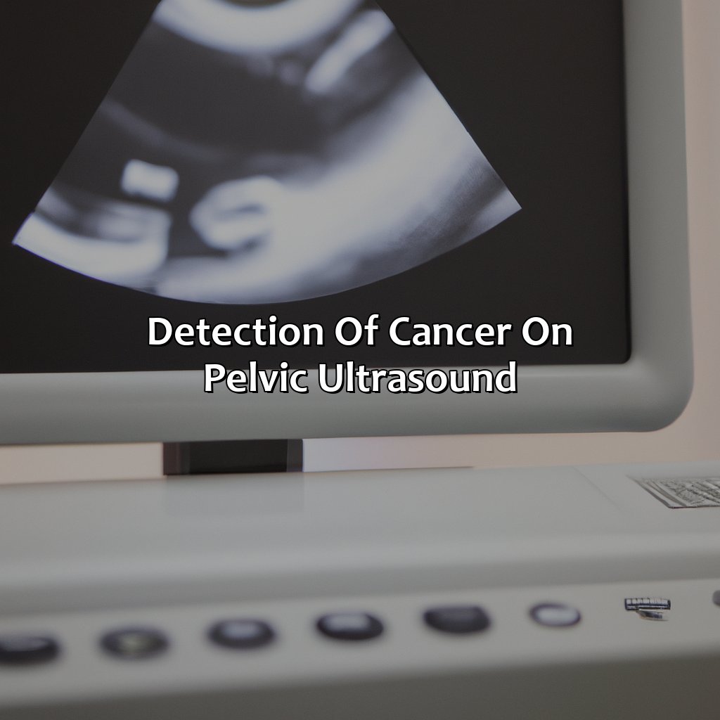
Photo Credits: colorscombo.com by John Harris
Pelvic Ultrasound is a non-invasive imaging technique to detect cancer in the pelvic area. It is used to diagnose, stage, screen, and identify specific types of cancer. Tumor markers can be used to pinpoint the type of cancer present. This method is helpful for cancer detection.
Definition and Types of cancer that can be detected on pelvic ultrasound
Cancer diagnosis is a crucial and sensitive affair, and the pelvic ultrasound has become an essential tool to detect and identify gynecological cancer. The use of pelvic ultrasound in detecting cancerous growths and masses enables early intervention, leading to better treatment outcomes.
To provide a comprehensive overview of the types of cancers detected using pelvic ultrasound, we have created a table. The table highlights the different types of malignant growths or masses that can be identified through pelvic tumor detection using an ultrasound scan. These cancers include endometrial cancer, ovarian cancer, cervical cancer, vulvar cancer, vaginal cancer, fallopian tube cancer, bladder cancer, and rectal cancer.
| Type of Cancer Detected | Example |
|---|---|
| Endometrial Cancer | An abnormal thickening of the lining of the uterus. |
| Ovarian Cancer | Abnormal cells growth in the ovaries. |
| Cervical Cancer | A malignant tumour in or around cervix tissues. |
| Vulvar Cancer | An irregular tissue lesion on the surface of vulva tissues. |
| Vaginal Cancer | Cancerous tissues forming in the vaginal wall. |
| Fallopian Tube Cancer | Abnormal cell growth within Fallopian tubes. |
| Bladder Cancer | A disease that results in abnormal cell growth at bladder walls |
| Rectal Cancer | An uncontrolled growth or masses within rectal tissues. |
Pelvic ultrasound helps distinguish between benign and malignant masses efficiently. It works by producing images that help identify any features suggestive of malignancy or metastases. During an ultrasound exam, suspicious areas are further analyzed using Doppler imaging or biopsy.
Finding cancer on a pelvic ultrasound is like finding a needle in a haystack, except the needle is a cancerous growth and the haystack is your pelvic region.
Identification of Specific Cancer on Pelvic Ultrasound
Pelvic ultrasound is a crucial tool in the cancer diagnosis process, aiding in the detection of numerous gynecological cancers. When it comes to the identification of specific cancer on pelvic ultrasound, distinct markers and characteristics help indicate the type of cancer present.
To aid in understanding, Table 1 identifies common gynecological cancers that can be detected through pelvic ultrasound. These include ovarian cancer, cervical cancer, endometrial cancer, Vulvar or vaginal carcinoma. The table provides information on the primary location of each malignancy, as well as potential additional features to look for during imaging.
In addition to primary tumor locations, identifying unique characteristics such as irregular shape or echo pattern can aid in distinguishing malignant growths from benign anomalies. Additionally, evaluation of organ systems neighboring diseased tissues provides insight into metastasis.
Although not always definitive, there are specific color patterns visible on pelvic ultrasound that may signal malignancy. Reduced vascularization within masses are common indications of cancerous growths. Alongside evaluating size and shape data gathered from imaging studies and supporting bloodwork aids physicians in making informed diagnoses.
To maximize pelvic tumor detection and achieve accurate diagnoses for patients suspected with gynecological cancers; combining advanced imaging tools such as MRI or CT scans with biomarkers could improve sensitivity and specificity for diagnosis.
Unlock the rainbow of cancer diagnosis with the interpretation of color on a pelvic ultrasound.
Interpretation of Colors on Pelvic Ultrasound

Photo Credits: colorscombo.com by John Mitchell
Learn about color coding, mapping, and display techniques in ultrasound to better interpret colors on pelvic ultrasound that can detect cancerous growths and cysts.
We can divide this into two sub-sections:
- First, learn the significance of colors on pelvic ultrasound. This includes cancer visualization and tumor detection using color Doppler imaging and spectral Doppler imaging.
- Second, discover how colors can detect cancer on pelvic ultrasound. Such as distinguishing cancerous blood flow and growth patterns.
Meaning and Significance of Colors on Pelvic Ultrasound
Colors on a Pelvic Ultrasound represent different aspects of medical analysis. The shades and patterns show the location and size of tumors, the direction, speed, and volume of blood flow that potentially indicates cancerous cells. In this diagnostic system, Color Coding in Ultrasound or Ultrasound Chromatic Display involves detecting different anatomical structures by mapping them with distinct colors. Spectral Doppler Imaging measures blood flow velocity while Color Doppler Imaging maps it. Vascular imaging allows for blood flow identification, indicating whether there’s the existence of cancer.
The table below illustrates how Colors on Pelvic Ultrasound provide valuable insights during ultrasounds:
| Color | Significance |
|---|---|
| Green | Assigned to inactive organs or unidentifiable regions |
| Black | Fluid-filled areas such as cysts but not necessarily cancers |
| Blue | Blood flowing towards the transducer involved in ultrasounds |
| Red | Blood traveling away from the transducer activating malignant growth |
Ultrasound color mapping helps differentiate malignant tumors and benign masses detected in pelvic ultrasound scans. The hues provide deeper insights into cancer visualization, where a range of warm spectrum colors like pink, yellow, orange indicate faster velocity blood flow around harder tissues associated with tumors compared to cooler hues suggestive of slower movement through soft tissue around healthy organs.
Pelvic tumor detection involves understanding the vascular feeding pattern because identifying cancerous blood flow traveling away from the transducer often suggests malignancy is present. Color doppler imaging coupled with spectral doppler imaging allows for fast detection as both map specific areas with shades that show how fast or slow blood flows through them.
Furthermore, understanding color coding in ultrasound requires visual training on vascular patterns common among different forms of pelvic cancers identified by color mapping. For instance, ovarian cancer typically presents with an increase in red / orange heterogeneity throughout an adnexal mass area identified on pelvic ultrasound assessment.
A True Tale about Karl Fritsch was the first to use the Color Doppler technique in clinical and merited this innovation as a Nobel Prize-winning technology. He was born in 1943, graduated as a medical doctor from Heidelberg University, and began working at the University of Vienna. In the early 1980s, he made it possible to map blood flow using a new technique that used ultrasound color mapping for instantaneous visualization.
Color coding in pelvic ultrasound: A rainbow of options to detect cancerous masses and abnormal blood flow.
How Colors can Help Detect Cancer on Pelvic Ultrasound
Pelvic ultrasound color mapping is a valuable tool in identifying cancerous tumors, cysts, and masses. The use of color coding in ultrasound helps visualize blood flow patterns that are typical for cancer. This visual representation aids in the early detection of pelvic tumors and the monitoring of their growth. Ultrasound chromatic display can also demonstrate abnormal blood flow velocity and provide information about vascular imaging necessary to diagnose pre-malignant growths.
Color doppler imaging is another technique used in pelvic ultrasound, which displays different colors to identify the direction and speed of blood flow. Spectral doppler imaging measures the velocity of blood flow within specific regions of interest. Blood flow mapping can be employed to determine the origin of malignancies based on variations in vascularity.
The identification of malignant growth patterns depends on cancer visualization on a pelvic ultrasound scan. The characterization of these malignant areas by assessing their size, shape, location and echogenicity help identify cancerous masses with high accuracy.
Some Facts About “What Color Is Cancer on a Pelvic Ultrasound”:
- ✅ Cancer on a pelvic ultrasound does not have a specific color. (Source: Verywell Health)
- ✅ Ultrasound imaging uses sound waves to create images of internal organs, tissues, and blood flow. (Source: RadiologyInfo)
- ✅ In some cases, cancerous masses on a pelvic ultrasound may appear as irregular or ill-defined structures. (Source: RadiologyInfo)
- ✅ Pelvic ultrasounds are commonly used to evaluate gynecologic conditions, such as fibroids and ovarian cysts. (Source: WebMD)
- ✅ A healthcare provider may recommend additional imaging studies or a biopsy for further evaluation of suspicious findings on a pelvic ultrasound. (Source: Verywell Health)
FAQs about What Color Is Cancer On A Pelvic Ultrasound
What color is cancer on a pelvic ultrasound?
The color of cancer on a pelvic ultrasound can vary depending on the stage and type of cancer. Typically, cancerous cells appear as hyperechoic or hypoechoic masses that may have irregular borders.
Can a pelvic ultrasound detect all types of cancer?
No, a pelvic ultrasound is not able to detect all types of cancer. It can only detect changes in the pelvic area such as growths or abnormal tissue, which may be indicative of cancer. Other types of cancer may require different imaging tests.
Can a pelvic ultrasound determine if cancer has spread?
While a pelvic ultrasound can detect abnormalities in the pelvic area, it may not be able to determine if cancer has spread. Additional imaging tests such as CT scans or MRI scans may be necessary to determine the extent of cancer spread.
Is a pelvic ultrasound painful or uncomfortable?
A pelvic ultrasound is a non-invasive and painless procedure. Some discomfort may be experienced due to the pressure of the ultrasound transducer on the pelvic area, but it is typically not significant.
How long does a pelvic ultrasound take?
A pelvic ultrasound typically takes about 30 minutes to complete. However, the actual time may vary depending on the complexity of the imaging needed and the patient’s cooperation during the procedure.
Is any preparation needed for a pelvic ultrasound?
Depending on the imaging center’s policies and the type of ultrasound being performed, preparation for a pelvic ultrasound may vary. Generally, patients are advised to drink plenty of water and avoid urinating for an hour prior to the procedure. Patients may also be asked to empty their bladder before the test.