Key takeaway:
- The human brain does not have a color, as it is made up of different structures and cells that have varying hues and shades. The perception of color in the brain comes from the activity of neurons and the way they process visual information.
- The anatomy of the brain also affects its color appearance, as different parts of the brain have different color representations. The cerebral cortex, for instance, may have different hues depending on its function and location.
- The presence of pigments in the brain, such as melanin and neuromelanin, may also contribute to its coloration. However, more research is needed to fully understand the role of pigments in the brain’s coloration and function.
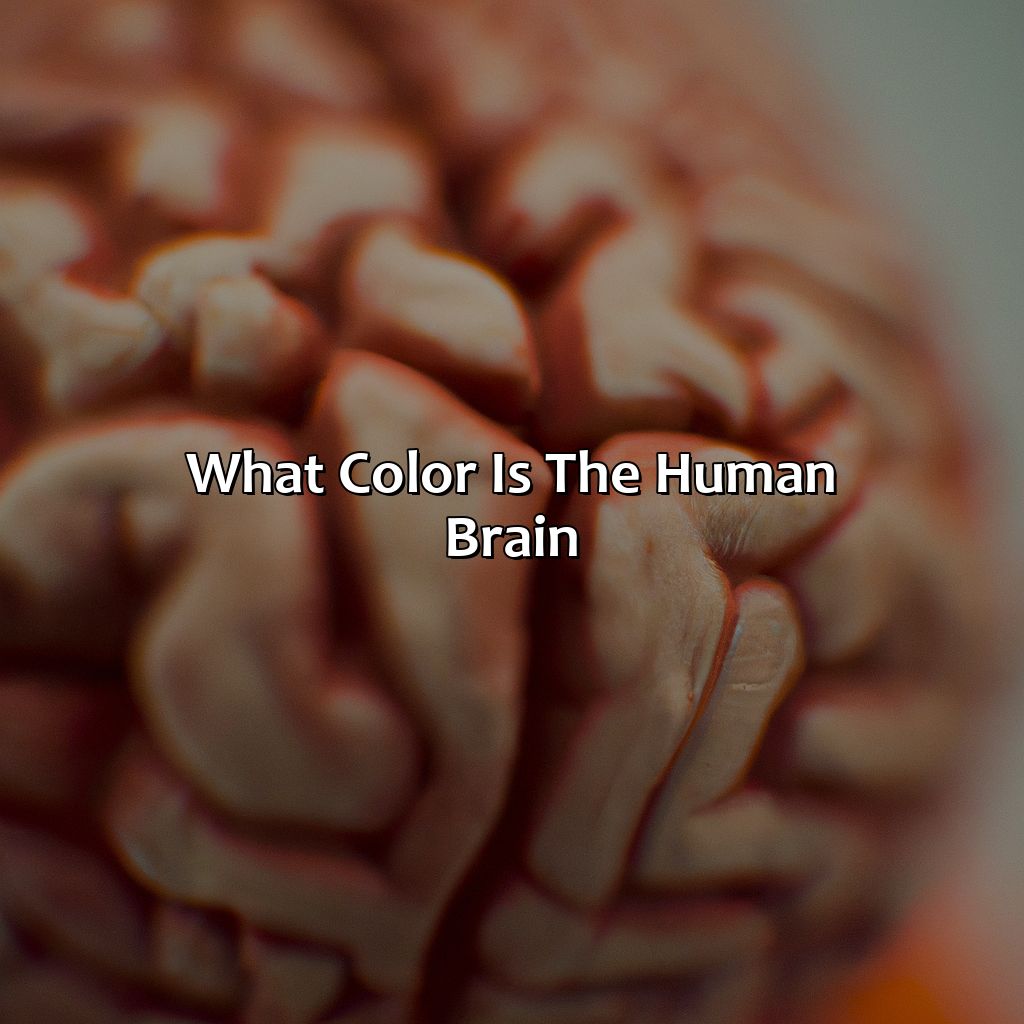
The Color of the Human Brain
Photo Credits: colorscombo.com by William Scott
The color of the human brain has been a topic of curiosity for many. While it may seem like an easy question, the answer is not as straightforward. The human brain lacks pigment, making it colorless. However, there are various factors that influence our perception of the brain’s color. The lighting in which it is viewed, as well as the surrounding colors, can create a perception of a specific color.
Research has suggested that certain colors can impact the brain’s functionality and mood. The color psychology of the brain is an area of study that explores how color affects our thoughts and behaviors. Different colors are associated with different emotions and can have a significant impact on our mental well-being.
The human brain color spectrum includes shades of white, grey, and some pinkish hues due to the presence of blood vessels. While the color of the brain itself may not be significant, brain color representation in media and popular culture can influence our understanding of it.
To enhance brain functionality and overall mental well-being, incorporating specific colors in our environment can be helpful. For example, blue has a calming effect and can aid in concentration, while red is associated with increased energy levels. Incorporating these colors into our surroundings can have a positive impact on our mood and productivity.
Understanding the perception and representation of brain color can provide valuable insights into the human brain’s functionality. Although the answer may not be as simple as we initially thought, exploring this topic can lead to a better understanding of how our brains work and how we can optimize our surroundings to enhance our mental health.
Understanding the Anatomy of the Human Brain
Photo Credits: colorscombo.com by Gabriel Green
To grasp the anatomy of the human brain, we’ll explore the colors of the cerebral cortex, grey matter, and white matter. This will help us comprehend the brain’s color coding. Two sections will be examined: the hemisphere colors, color symbolism in neuroscience, and the color of the cerebellum.
The Cortex
The Outer Layer of the Human Brain
The outer layer of the human brain, responsible for conscious decision-making and thought processes, has been a topic of interest in both neuroscience and psychology. This region is known as the cerebral cortex and is divided into four main lobes: frontal, parietal, temporal, and occipital.
This area contains more than 16 billion neurons that communicate with each other to create our thoughts, emotions, and behaviors. It is important to note that the color symbolism in neuroscience has been an evolving subject with the advancement in imaging techniques. Interestingly, there have been studies that suggest possible brain hemisphere colors but no clear scientific consensus exists.
Ready to get your brain in gear? Let’s dive into the fascinating world of the cerebellum.
The Cerebellum
The cerebellum is a part of the brain located in the posterior cranial fossa. It is responsible for coordinating movements, maintaining posture and balance, as well as assisting in cognitive functions such as attention and language processing. The cerebellum contains several layers of cells, including granular cells and Purkinje cells that contribute to its complex neural circuitry. The cerebellum receives input from multiple sensory systems and modulates output to various motor areas. It is composed of over half of the neurons in the brain but only accounts for approximately 10% of its total volume.
Unique details about the cerebellum include its ability to learn and adapt to changes in sensory input through a process called plasticity. This allows it to continually fine-tune motor movements throughout life. Another unique feature is its role in regulating emotional responses, with studies suggesting that damage or dysfunction of the cerebellum can be linked to psychiatric disorders such as bipolar disorder.
Explore further research on the exciting nature of this fascinating organ! Don’t miss out on understanding more about this essential aspect of our brain’s composition and function.
Seems like the human brain not only has a complex anatomy, but a colorful personality too!
Pigment in the Human Brain
Photo Credits: colorscombo.com by Henry Lewis
To understand how the brain gets its color, look into melanin and neuromelanin, hemoglobin and iron. These components offer an answer to the puzzle. Investigate the brain color theory, what the colors mean, and the neuroscience behind color and emotions. Uncover how color affects and shapes the workings of the brain.
Melanin and Neuromelanin
Melanin and neuromelanin are color pigments present in the human brain. Melanin is mainly associated with skin color, but it is also found in the brain where it provides neuroprotection against oxidative stress. Neuromelanin, on the other hand, is a byproduct of dopamine metabolism and is primarily found in specific neuronal populations such as the substantia nigra and locus ceruleus.
The role of melanin and neuromelanin extends beyond their pigment properties and can also influence neurotransmitter signaling, cytokine production, and synaptic plasticity. High levels of neuromelanin are observed in certain regions responsible for movement control, while low levels in areas such as the prefrontal cortex.
Interestingly, both melanin and neuromelanin have been linked to age-related neurodegenerative disorders like Parkinson’s disease. In Parkinson’s patients, there is a significant loss of cells that contain neuromelanin leading to abnormal movement behaviors.
It is worth noting that due to their non-reflective pigment properties, melanin and neuromelanin do not alter conventional imaging techniques’ sensitivity. However, specialized imaging methodologies like MR Spectroscopy can detect these pigments’ specific signals.
In summary, melanin and neuromelanin play crucial roles beyond providing coloration to the human brain. Researchers continue to explore these pigments’ protective functions further to deduce their potential significance or implication in various brain functions or pathological conditions.
Looks like the human brain is not just gray matter, but also a touch of iron and a little bit of blood for that extra oomph.
Hemoglobin and Iron
Iron and hemoglobin are essential to the functioning of the human brain as they aid in oxygen transportation. Hemoglobin is a protein found in red blood cells responsible for binding oxygen and transporting it throughout the body. Iron is a crucial component of hemoglobin, playing a vital role in its function. Without adequate iron and hemoglobin levels, brain tissue can experience hypoxia, leading to cognitive impairment or dysfunction.
In addition to their important role in oxygenation, recent studies have suggested potential neuroprotective effects of both iron and hemoglobin. For example, iron has been shown to have antioxidant properties that may help protect the brain from oxidative stress, while hemoglobin has been implicated in reducing damage caused by traumatic brain injuries.
Interestingly, abnormal levels of iron and hemoglobin have also been linked to various neurological disorders such as Alzheimer’s and Parkinson’s diseases. The accumulation of excess iron in specific regions of the brain has been associated with the development of these diseases.
A study conducted by Rajesh Kumar et al., published in 2019, revealed high levels of ferritin (a protein containing iron) in Parkinson’s patients using Magnetic Resonance Imaging. Hence it can be inferred that Hemoglobin and Iron play significant roles inside our brains along with having neuroprotective effects when at healthy levels.
Peek into the colorful world of the brain with neuroimaging techniques and discover the fascinating hues of brain color map and coding in neurological research.
Imaging Techniques for Observing Brain Color
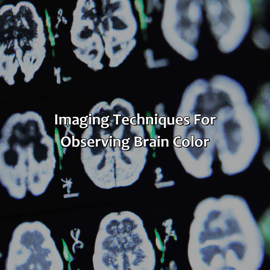
Photo Credits: colorscombo.com by Kenneth Flores
Observing brain color more accurately requires imaging techniques. Two of these are Magnetic Resonance Imaging (MRI) and Diffusion Tensor Imaging (DTI). Color coding helps study brain color psychology and sensitivity. We’ll discuss these imaging techniques and their importance for neurological research.
Magnetic Resonance Imaging
Magnetic resonance imaging (MRI) is a technique used to generate high-resolution images of the human brain. By creating a strong magnetic field around the patient and then using radio waves, MRI can detect differences between tissue types within the brain. These signals are then processed by computer algorithms to produce detailed images of the brain’s structures.
In clinical settings, MRI is often used to diagnose neurological disorders such as tumors and multiple sclerosis. It can also be used to monitor changes in the brain over time, such as during the progression of Alzheimer’s disease.
Notably, there are several variations of MRI available, including functional MRI (fMRI) which measures changes in blood flow that occur during neuron activity. DTI-MRI adds directional data about water diffusion and provides information on structural connectivity between different regions of interest.
To optimize MRI results, patients should remain still during imaging and avoid any metal objects or clothing that may interfere with the magnetic fields. Doctors will typically use contrast agents to highlight specific areas of the brain for additional detail.
Overall, Magnetic resonance imaging remains a valuable tool for researchers exploring the anatomy and functionality of the human brain.
“The brain may be grey, but with DTI we can see it in a whole new light.”
Diffusion Tensor Imaging
Here is a table showing some technical specifications for DTI:
| Resolution | Typical voxel size: 2-3 mm isotropic |
| Sequences | Single or multi-shell acquisition |
| Sensitivity | High sensitivity to white matter microstructure |
| Applications | Detecting brain abnormalities and characterizing neural architecture |
DTI provides important information about structural connections in the brain that cannot be detected using traditional MRI. Through DTI, researchers can study disorders such as Alzheimer’s and schizophrenia that involve abnormal communication between various regions of the brain.
Interestingly, DTI has also been applied in areas outside of neuroscience, such as engineering, psychology, and even education. For example, engineers have used DTI to study how water flows through concrete pavement.
A true fact about DTI: “DTI was first introduced in the early 1990s by Peter Basser.” (source: NIH)
The only thing scarier than abnormal brain coloration in fiction is the actual science behind it.
Abnormal Brain Coloration and Associated Diseases
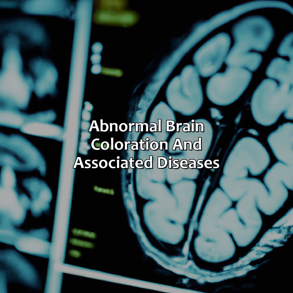
Photo Credits: colorscombo.com by Larry Clark
Discover the science of brain color and strange hues in the human mind. Delve into Albinism, Parkinson’s Disease and more. Explore the research areas within this, and the colors that exist in fiction. Go further than physicality and understand the energy and data flow of the brain.
Albinism
Individuals with albinism often have pale or white hair and skin due to the absence of melanin. They may also have lighter eye colors than those without albinism, such as blue or pale green eyes. The condition can occur in all races and ethnicities but is more prevalent in some populations.
Interestingly, while people with albinism do not produce enough melanin for their skin or hair color, they still produce normal levels of neuromelanin. Neuromelanin is responsible for pigmenting neurons in specific regions of the brain.
Historically, individuals with albinism have been subject to discrimination and ostracization in some cultures due to negative cultural beliefs about the condition. However, education and awareness efforts have helped bring attention to these harmful attitudes towards individuals with albinism.
Looks like Parkinson’s isn’t just shaking things up, it’s changing the color of your brain too.
Parkinson’s Disease
Parkinson’s disease is a neurodegenerative disorder that affects movement control and mental function. It is caused by a decrease in dopamine-producing cells in the brainstem. The reduction of dopamine leads to muscle rigidity, tremors, and difficulty initiating movements.
Parkinson’s is known for its motor symptoms, but it also has non-motor symptoms such as cognitive decline and depression. Treatments are available to manage symptoms, including medications that increase dopamine levels or improve symptom relief.
Research suggests that Parkinson’s disease may have underlying genetic factors and that environmental toxins may contribute to its development. More studies are needed to understand the complexities of this challenging disease.
Pro Tip: Early diagnosis can help improve quality of life and better manage symptoms. Regular exercise has also been shown to be beneficial in slowing the progression of Parkinson’s disease.
Some Facts About the Human Brain’s Color:
- ✅ The human brain is not actually gray, but rather a pinkish-beige color. (Source: Live Science)
- ✅ The pigmentation of the brain comes from the mix of blood vessels and neurons. (Source: BrainFacts.org)
- ✅ The color of the brain can change due to certain neurological conditions, such as Parkinson’s disease. (Source: Healthline)
- ✅ During fetal development, the brain is typically black because it has not yet developed pigmentation. (Source: ScienceDirect)
- ✅ Although the brain is not gray, the popular image of the gray brain persists due to medical imaging techniques that use grayscale. (Source: The Conversation)
FAQs about What Color Is The Human Brain
What color is the human brain?
The human brain is not technically a color, as it does not have pigment. However, it is often portrayed as gray or pinkish-gray in medical illustrations.
Is the color of the human brain the same for everyone?
Yes, the color of the human brain is the same for everyone. However, due to variations in lighting and visualization techniques, it may appear slightly different in different images.
Does the color of the brain change during development or with age?
The color of the brain does not change significantly during development or with age. However, certain medical conditions or injuries can cause changes in the appearance of the brain on imaging studies.
What causes the grayish color of the brain?
The grayish color of the brain is due to a type of brain tissue called gray matter. Gray matter contains nerve cell bodies, which play a key role in information processing and decision-making.
Why do some people say the brain is pinkish in color?
Some people may describe the brain as pinkish in color because it contains blood vessels, which can give it a slightly reddish or pinkish tint. However, this is not the dominant color of the brain.
Does the color of the brain have any significance for health or disease?
The color of the brain itself does not have specific significance for health or disease. However, changes in the appearance of the brain on imaging studies can help diagnose and monitor medical conditions affecting the brain.